|
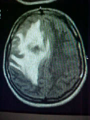
| FLAIR Axial MR Brain at 32 weeks of pregnancy |
|
| shows hemorrhagic infarct in the right centrum semiovale with extensive peri-lesional edema |
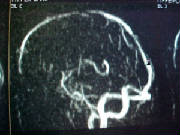
| Sagittal view of MR Venogram Brain |
|
| shows involvement of straight sinus and other deep venous system |
In the pictures above you see MR images. One is a FLAIR
Axial MR image and the other a sagittal section of MR venogram of a patient who presented in the 32nd week of her pregnancy
with a left hemiparesis. The MR showed a hemorrhagic infarct in the deep white matter of the right cerebral hemisphere with a
large peri-lesional edema. The MRV confirmed the clinical suspicion of Cerebral Sino-venous thrombosis. At our center ( Apollo
Hospitals, Hyderabad ), we now have a large series of such patients, and possibly we have the largest experience in thrombolysis
of CSVT. In this patient, thrombolysis would have been risky for the mother as well as the baby, and the gyneocologist wanted
to delay delivery till at least the 36-38th week. We tried to decrease the edema with the help of mannitol and steroid, which
would also help in maturation of the fetal lung. she returned by the 36th week with seizures, which were slightly difficult
to control. A planned cesarian section was done at the 36th week, and both mother and child are doing well. We have started
her on oral warfarin and she is doing very well. She will need anti-coagulation for at least another 6 months, and we will
have to reassess the need for further anti-coagulation then. We were happy to see a happy ending for the pregnancy after such
a devastating event in late pregnancy.
We have to analyse all our patients and come to some sort of understanding
about the role of thrombolysis in these patients. Though in the acute setting many patients do well even without it, some
of the more chronic problems cannot be avoided if it is not done.
| FLAIR Axial MR image of Brain at 32 weeks |
|
| Shows hemorrhagic infarct right centrum semiovale with extensive peri-lesional edema |
This text will describe the picture above.
If someone other than me has written an article, I'll be sure to include a byline at the bottom.
This article contributed by Jane Turner.
|


